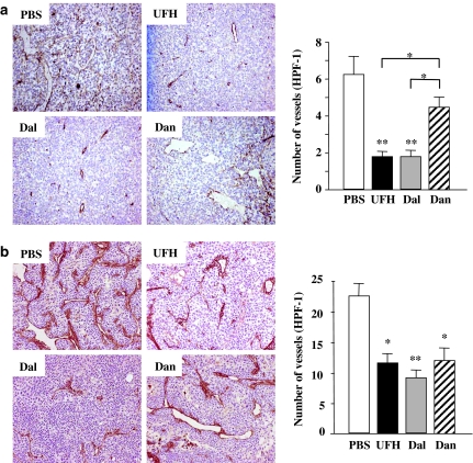
Figure 2.
(a) Immunohistochemical staining for factor VIII-related antigen of the LLC tumours. Left: representative photographs from each group are shown (magnification, × 100). Right: the effects of the PSAs on the tumour microvessel densities. Each value is the mean±s.e. (n=5–7 per group) of vessel counts observed with a high-power field (magnification, × 200). *P<0.05 among PSAs by one-way ANOVA with Fisher's least-significant-difference test. **P<0.01 compared with PBS group by Student's unpaired t-test. (b) Immunohistochemical staining for factor VIII-related antigen of KLN205 tumours. Left: representative photographs from each group are shown (magnification, × 200). Right: the effects of the PSAs on the tumour microvessel densities. Each value is the mean±s.e. (n=5–7 per group) of vessel counts observed with a high-power field (magnification, × 200). *P<0.05, **P<0.01 compared with the PBS group by the Student's unpaired t-test. There was no significant difference between the PSAs by one-way ANOVA with Fisher's least-significant-difference test.

