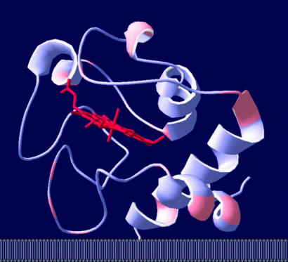
Fig. 5.
Predicted orientation of cytochrome c on the inner mitochondrial membrane at 100 mM KCl. The heme group is shown in red. Lysine residues shown in pink may interact with the negatively charged residues on the subunits of complex III/complex IV in addition to the head groups of cardiolipin. Further details are described in Results.

