Figure 2.

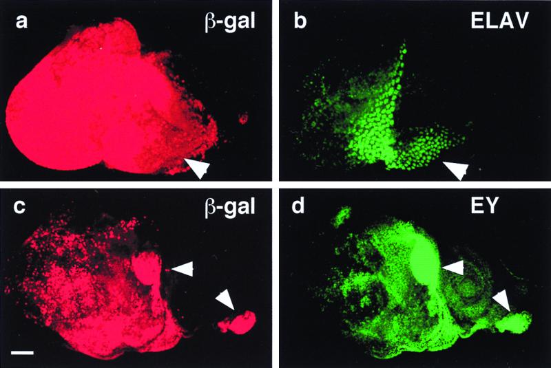
Induction of ectopic photoreceptor cells and eyeless by Notch signaling. (a) Anti-β-galactosidase (β-gal) antibody staining of a UAS-Nact UAS-lacZ ey-GAL4 eye-antennal disc. Activation of the lacZ reporter reflects the distribution of constitutively activated Notch protein. Arrowhead indicates hyperplastic portion. (b) Immunostaining of same disc as in a with antibody against the neuronal marker ELAV. In the hyperplastic portion (arrowhead) ectopically induced photoreceptors. (c) Anti-β-galactosidase antibody staining of a UAS-Nact UAS-lacZ ey-GAL4 eye antennal disc. Activation of the lacZ reporter reflects the distribution of Nact protein. Arrowheads indicate areas of strong lacZ expression. (Bar indicates 50 μm.) (d) Immunostaining of same disc as in c with antibody against EY. Ectopic EY expression is induced in the areas of strong lacZ expression (arrowheads).
