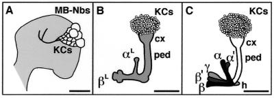
Figure 1.
Development of the MBs. (A) Embryonic MB primordium. Lateral image of an embryonic brain hemisphere at stage 17 showing the four MB neuroblasts (MB-NBs) that are located at the anterior end (in neuraxis) of the brain and give rise to the embryonic Kenyon cells (KCs). Axonal tracts of the peduncle and two orthogonal lobes are pioneered by this stage. (Bar = 10 μm.) (B) Larval MB. The larval MB structure is basically an extension of the embryonic one with increasing numbers of Kenyon cells (KCs) and their axons. The peduncle (ped) split into the dorsal (αL) and medial (βL) lobes. cx, calyx. Third instar. (Bar = 50 μm.) (C) Late pupal and adult MBs. After massive reorganization during the first half of the pupal stage, two dorsal (α and α′) and three medial lobes (β, β′, and γ) are formed. Together with the heel (h), these adult-type structures can be classified into three axonal projection groups (α/β, α′/β′, and γ/heel), which are indicated with different shadings. (Bar = 60 μm.)

