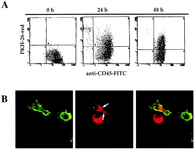
Figure 3.
Uptake of apoptotic bodies by DCs during differentiation. DCs, at day 2 of culture, were cocultivated with irradiated M3-CSM cells, previously labeled with red fluorescent dye PKH-26. Flow cytometry analysis (A) for PKH-26-red and CD45-FITC expression by DCs was performed after 24 and 48 h of cocultivation. Confocal microscopy analysis (B) showed the presence of intracellular apoptotic bodies (red staining) within the cytoplasm of DCs labeled with a HLA-DR-FITC mAb (green staining).

