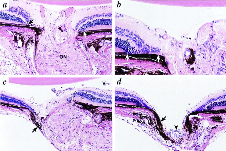
Figure 2.
Colobomas in 6-mo-old Bst/+ mice. (a) In a minimally involved optic nerve, the retina and RPE terminate prematurely before reaching the margin of the optic nerve (arrow). The RPE (white arrow) ends before the nerve on the opposite side. Nerve fibers entering the nerve are not clearly identified, although the optic nerve (ON) demonstrates nearly normal architecture. Original magnification, ×400. (b) At the edge of the optic disc, there is folding of the retina with direct contact between the choroid and inner and outer nuclear layers (white arrows). (c) In more severely affected mice, there is distortion of the peripapillary retina, RPE, and choroid. Misplaced choroidal melanocytes are present in the periphery of the optic nerve (arrow). Neither lamina cribrosa nor nerve fibers are identified. Persistent hyaloid vessels (V) are present in the posterior vitreous. (d) The optic nerve is not identifiable, other than by a tiny cluster of undifferentiated cells (arrowhead). Choroidal pigment (arrow) prolapses into the empty cup. Original magnification in b–d, ×200.

