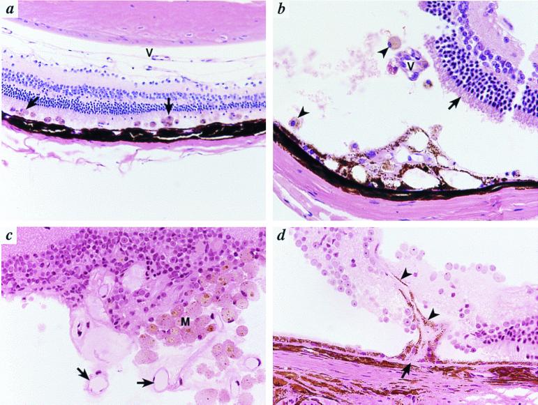
Figure 4.
Retinal detachment and SRN in Bst/+ mice. (a) There is a flat detachment of the retina containing many pigment-filled macrophages (arrows). Persistent hyaloid vessels (V) are present. Original magnification, ×200. (b) There is a more extensive retinal detachment with loss of photoreceptor outer segments (arrow). Pigmented macrophages are present (arrowheads). There is a focus of fibrovascular proliferation involving the RPE (*) that contains eosinophilic connective tissue and small vascular channels. Additional vascular channels (V) that appear to connect to this focus in other planes of section were also present. The tears in the retina are the result of artifacts of sectioning. (c) In an extensive retinal detachment, numerous proliferating vessels (arrows) and macrophages (M) are evident. (d) A new vessel arises from the choriocapillaris (arrow) and extends into the subretinal space and retina (arrowheads). Original magnification in b–d, ×400.

