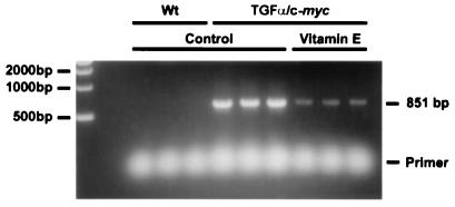
Figure 6.
PCR detection of deletion in liver mtDNA. Total hepatic DNA samples from 10-wk-old wt, and TGFα/c-myc mice fed with control or VE-supplemented diet were subjected to PCR amplification and separated using a 1% Tris-acetate-EDTA-agarose gel electrophoresis as described in Materials and Methods. The position of PCR deletion product of 851 bp is indicated. The deletion was undetectable in 10-wk-old wt livers in the absence of oxidative stress. In contrast, the deletion was readily detectable in the age-matched TGFα/c-myc mice. The deletion product was more abundant in TGFα/c-myc mice fed with control diet, which manifest increased ROS generation.

