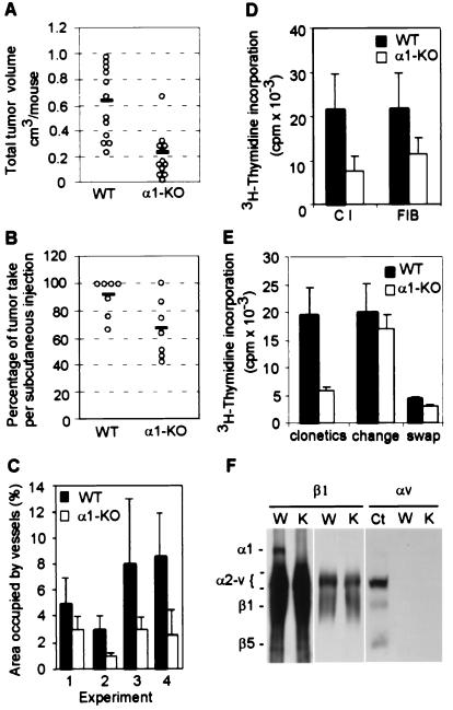
Figure 2.
(A) Tumor volume in wild-type (WT) and α1-null (α1-KO) hosts at 14 days. ○ indicate the total tumor volume for each mouse and the solid line the mean of samples. Four independent experiments were performed, each with a minimum of three animals per genotype. α1-null hosts show a significant reduction in tumor volume (P < 0.05) (B) Proportion of tumors growing from the initiating cell bolus after 10–14 days. The number of tumors grown from the injected initiating cell bolus of 105 cells was counted for each animal. ○ indicate the mean result for each experiment, and the solid line the mean of means. Seven independent experiments were performed, each with a minimum of three animals per genotype. α1-null hosts show a significant reduction in tumor take (P < 0.05). Although tumor size increased with growth time, percentage take did not change with growth time. (C) Tumors from α1-null hosts show reduced vascularity as measured by extent of CD31 positive structures (P < 0.05). Bars and errors indicate the mean and SD (five tumors per genotype were examined in experiment 1, three per genotype in experiment 2, seven per genotype in experiment 3, five per genotype in experiment 4). (D) α1-Null primary lung endothelial cells show reduced proliferation on collagen and fibrinogen as compared with wild-type cella (P < 0.05). The y axis indicates absolute cpm incorporation of primary endothelial cells grown as described in Materials and Methods. Bars and errors indicate the mean and SD of three different experiments (with a total of six animals for each group). (E) Inhibition of proliferation of α1-null endothelial cells is the result of a soluble factor. The y axis is the same as in D. Means and SD are of triplicate samples of endothelial cells from three wild-type and α1-deficient mice. α1-Null cells grow less than their wild-type counterparts when cultured in complete medium (“clonetics”). Changing the medium at 12 hourly intervals (“change”) rescues α1-null cell growth, returning growth to wild-type levels; while adding conditioned media from α1-null cells (“swap”) prevents wild-type cell growth. (F) Immunoprecipitation from lysates of wild-type (W) and α1-null (K) endothelial cells with anti-β1 or anti-αv antibody. The designation α2-v indicates the positions of subunits α2, 3, 4, 5, and v, which are incompletely resolved from one another. Anti-β1 antibody failed to precipitate α1 integrin in α1-null endothelial cells (Left). A light exposure of the same membrane is shown to reveal no differences in α2-v bands between wild-type and α1-null endothelial cells (Middle). Anti-αv antibody failed to precipitate αv integrin in lysates of endothelial cells of both genotype, unlike in those of control embryonic fibroblasts (Ct) (Right).

