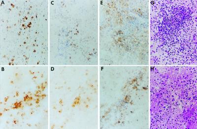
Figure 5.
Immunohistologic identification of inflammatory cell infiltrates. A/J female mice were injected intracerebrally with Neuro-2a cells (1 × 10⁵/5 μl) and 5 days later were injected intratumorally with 1 × 10⁷ pfu of HSV R3659 (A, C, E, and G) or HSV M002 (B, D, F, and H). After 6 days, the mice were killed, and their brains were removed intact and embedded in OCT for preparation of frozen sections. Serial 10-μm-thick sections were reacted with rat monoclonal antibodies to CD4+ (A and B) or CD8+ T (C and D) cells or macrophages (E and F); antibody binding was detected by using horseradish peroxidase-labeled anti-rat antibody, and sections were counterstained with Mayer's hematoxylin. Hematoxylin-eosin stained adjacent sections are also shown (G and H).

