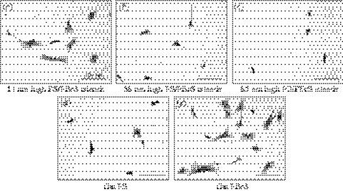
Figure 4.
Coomassie blue stained hFOB cells cultured for 3 h on nanoislands and flat substrata. Original 720×960 μm2 size images were taken at a 10× objective magnification and used for image analysis of cell dimensions. Cropped sample images (360×480 μm2) are shown with substrata notation and scale bar.

