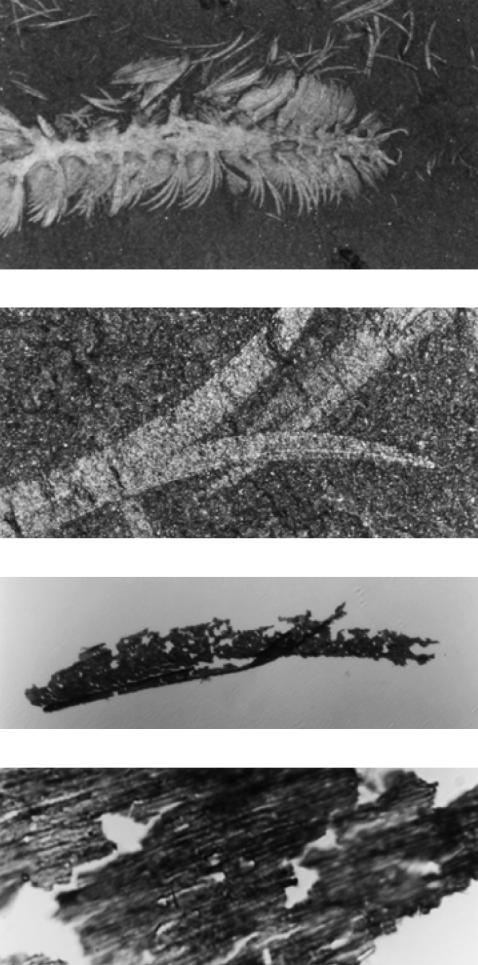
Figure 2.

Micrographs of the Burgess stem-group polychaete Canadia spinosa at increasing magnification—from ×10 to ×4000. The top picture shows the anterior half of the animal, the middle pictures show details of paleae (spines). The bottom picture shows the surface of a palea as removed from the rock matrix, revealing the remnants of a diffraction grating with a ridge spacing of 900 nm.
