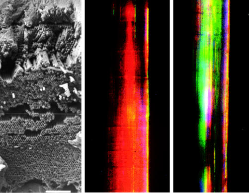
Figure 23.
Left: scanning electron micrograph of a ‘photonic crystal’ in the sea mouse Aphrodita sp. (Polychaeta). This shows a cross-section through the wall of a tube, or spine, constructed of nano-tubes, close-packed hexagonally. Internal diameters of the individual nano-tubes increase systematically with depth in the stack. Scale bar, 8 μm. Centre and right: light micrographs of a section of the spine showing the different colours obtained when the orientation in the horizontal plane varies (by 90°) with respect to the direction of the light source.

