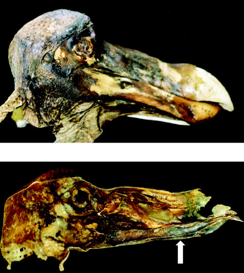
Figure 29.

The head of the Oxford dodo specimen. Above: right, lateral (external) view of the skin attached to the skull (note that the end of the bill is bare). Below: medial (internal) view of the skin from the left side of the head, without the skull (note the blue colouration near the end of the lower bill region, arrowed).
