
Figure 8.
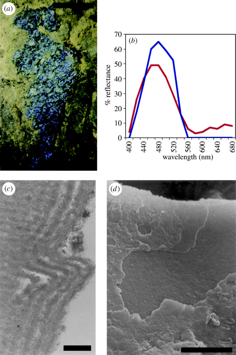
Elytron of an unidentified beetle from Messel, 50 Ma. (a) Light photograph of the specimen examined, embedded in rock. (b) Graphs showing the experimental optical (blue, solid line) and predicted (red, dashed line) reflectance from the elytron. (c) Transmission electron micrograph of the outer surface of a 60‐nm‐wide section of elytron, showing the fine lamination (running vertically in the picture) in a cross-section. The surface of the elytron has become wrinkled; the bend shown here represents a wrinkle. ‘Horizontal’ lines are an artefact of cutting. (d) Scanning electron micrograph of the outer surface of the elytron, perpendicular view, showing internal fine layers where outer layers are broken. Scale bars, a, 1 mm; c, 0.5 μm; d, 5 μm. From Parker & McKenzie (2003).