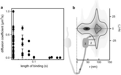
FIGURE 8.
Movement of HBV core capsids around NPC observed by dual-color wide-field microscopy. (a) Diffusion coefficients versus duration of HBV core capsid-NPC interactions. (b) Probability density distribution for the position of the capsid binding events with respect to the central axis of NPCs in polar coordinates r and φ determined from 23 binding events that lasted at least 2 frames. Distance between contours, 1′10−4. A sketch of the NPC (see Fahrenkrog et al. (29)) is aligned to the probability density plot. The highest probability for capsid binding is found for distances of 44 ± 9 nm from the central axis of an NPC. Bindings at larger distances occur with much lower probability. 1, cytoplasmic filaments; 2, luminal domain; 3, ring moiety; 4, nuclear basket.

