Fig. 1.
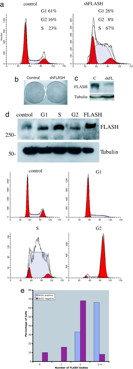
Down-regulation of FLASH results in S-phase block. (a) Cell-cycle distribution of HeLa cells transfected with GFP-spectrin and either pSUPER-scrambled (control) or pSUPER-FLASH-1 (shFLASH). Cells were stained with PI, and GFP-positive cells were analyzed by flow cytometry for DNA content. Percentages of cells in G1, S, or G2/M calculated by using ModFit program are indicated. Identical results were obtained by using pSUPER-FLASH-2 (data not shown). (b) Colony-forming assay of SAOS-2 cells transfected with pBabe-puro and either pSUPER-scrambled (control) or pSUPER-FLASH-1 (shFLASH), selected for 2 weeks with 1 μg/ml puromycin. Cells transfected with pSUPER-FLASH-1 show a large reduction in colony numbers. (c) Western blot showing that transfection of MCF-7 cells with pSUPER-FLASH-1 (shFL), but not with a scrambled vector (control), results in reduction of FLASH protein levels. (d Upper) Western blot showing FLASH expression in MCF-7 cells untreated (control) or treated with either 2 mM thymidine for 16 h (G1), followed by 4-h release in 24 μM deoxycytidine (S), or treated with 50 ng/ml nocodazole for 16 h (G2). As a positive control, cells transfected with GFP-FLASH (FLASH) were loaded. The Western blot was reprobed with tubulin to show equal loading. (d Lower) Cell-cycle distribution of an aliquot of cells used for the Western blot. (e) Percentage of IMR90 cells containing 0, 1, 2, or 3–4 FLASH bodies positive (blue) or negative (red) for BrdU incorporation. The number of FLASH bodies per cell increases in S phase cells as reported for CBs.

