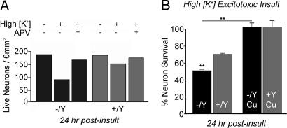
Fig. 5.
Mobr hippocampal neurons display increased sensitivity to excitotoxicity. (A) Representative experiment in which neurons were stimulated in high K+ buffer for 5 min, rinsed in ECS with 2 mM MgCl2, and then incubated in conditioned media for 24 hr after insult (High K+). Neurons were then assayed for viability as described in Materials and Methods. In some experiments, neurons were treated as described above in the presence of the NMDA receptor antagonist APV (where indicated). (B) Percent survival across all experiments was calculated from the ratio of live neurons on experimentally treated coverslips to live neurons on mock-treated coverslips from the same dish of plated coverslips. In some experiments, neurons were treated with 200 μM CuCl2 during the insult (Cu), which increased neuronal survival rates in both the +/Y and −/Y genotypes. (∗∗, P < 0.001.)

