Figure 3.
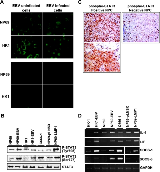
Activation of STAT pathways in EBV-infected nasopharyngeal epithelial cells. (A) Immunofluorescent staining with STAT3-specific antibody (original magnification, x100; upper panel). Lower panel shows negative controls in which no primary antibody was added. (B) Western blot analysis of STAT3 activation using phosphorylation-specific antibodies. Actin was included as loading control. (C) Immunohistochemical staining for phospo-STAT3. Left: Positive staining of phospho-STAT3 was exhibited in two primary NPC samples (original magnification, x400). Right: A case of NPC showed negative staining of phospho-STAT3. Positive staining of lymphocytes as internal positive control (original magnification, x400). (D) Expression of STAT3-associated genes in EBV-infected nasopharyngeal epithelial cells. Semiquantitative RT-PCR analysis of IL-6, LIF, SOCS-1, and SOCS-3.

