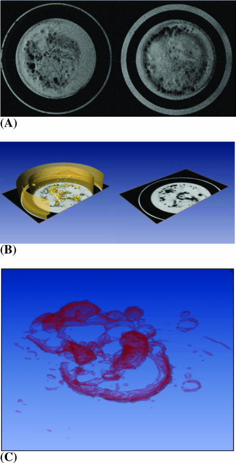
Figure 3.
(A) Representative axial MR images of two contiguous 0.5-mm-thick slices containing HUVECs labeled with 9 µg/ml Feridex. A lumen-like network formation of ECs 24 hours postseeding is apparent in these images, which were obtained using a T2-weighted spin-echo sequence with a field-of-view = 1.6 cm, acquisition matrix = 256 x 256, TE = 60 milliseconds, TR = 617 milliseconds, and number of averages = 2. (B) A three-dimensional reconstructed image of multislice data showing a lumen-like HUVEC network in the ECM gel (left). Orthogonal slices in the axial direction provide the ability to dynamically follow migration, invasion, and network structure through the ECM gel (right). (C) A three-dimensional reconstructed image from a multislice MR data set, with volume rendering demonstrating an intricate HUVEC network.

