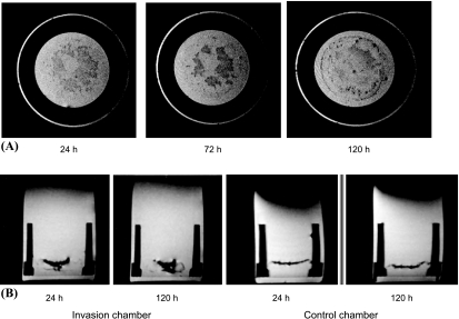
Figure 4.
(A) A panel of three axial MR images acquired 24, 72, and 120 hours postseeding. An increased number of HUVECs were detected with time in the layer containing MDA-MB-231 cancer cells, as evident from hypointense regions in this layer. HUVECs were labeled with 9 µg/ml Feridex. The MR images were obtained using a T2-weighted spin-echo sequence with field-of-view = 1.6 cm, acquisition matrix = 256 x 256, eight slices of slice thickness = 0.5 mm, TE = 60 milliseconds, TR = 617 milliseconds, and number of averages = 2. (B) Representative coronal MR images of a chamber containing MDA-MB-231 cells demonstrating the presence of Feridex-labeled HUVECs confined mainly to the upper seed layer 24 hours postseeding (i), with an increased presence of HUVECs in the cancer cell layer 120 hours postseeding (ii). By comparison, the control chambers showed no invasion of the ECM gel by the HUVECs, 24 hours (iii) and 120 hours postseeding (iv). The images were obtained in a single scan, using a T2-weighted spin-echo sequence with a field-of-view = 1.6 cm, eight slices of slice thickness = 1 mm, acquisition matrix = 256 x 256, TE = 60 milliseconds, and TR = 617 milliseconds.

