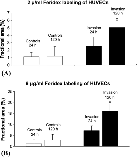
Figure 5.
Fractional area occupied by HUVECs in the cancer cell seed layer for (A) HUVECs labeled with 2 µg/ml Feridex and (B) HUVECs labeled with 9 µg/ml Feridex. The fractional area was calculated for T2-weighted MR images acquired with TE = 60 milliseconds and TR = 617 milliseconds. A significant increase (P < .05) was detected in the fractional area (mean ± SD) occupied by HUVECs in the cancer cell layer for chambers containing MDA-MB-231 breast cancer cells (n = 6 for 2 µg/ml Feridex; n = 5 for 9 µg/ml Feridex), whereas no significant change (P < .05) was observed in the corresponding layer in the absence of cancer cells (n = 6 for 2 µg/ml Feridex; n = 4 for 9 µg/ml Feridex).

