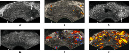
Figure 2.
A 75-year-old man with prostate-specific antigen of 5.1 and Gleason 8 cancer in the left base: transverse images through the base of the prostate. A. Conventional gray scale image shows a hypoechoic area in the left base (arrows). B. Power Doppler image shows no significant increase in flow in this hypoechoic area. C. Harmonic gray scale during contrast infusion shows a clearly defined area of focal enhancement, corresponding to the cancer (arrows). D. Harmonic gray scale with intermittent imaging shows a less well-defined, larger area of parenchymal enhancement around the cancer. E. Contrast-enhanced color Doppler image shows increased flow associated with the cancer. F. Contrast-enhanced power Doppler image also shows increased flow associated with the cancer.

