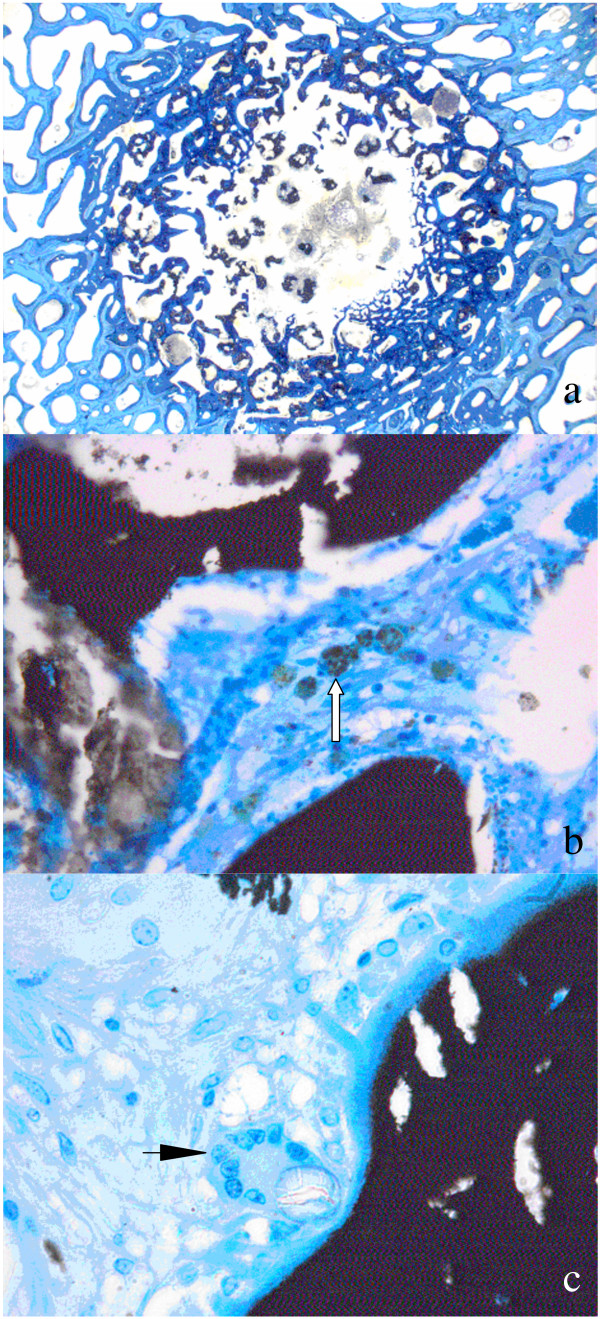
Figure 11.
Histology samples: While ground sections (30–40 μm, surface stained with toluidine blue) are well suited for the assessment of osseointegration, new bone formation, materials resorption and histomorphometrical measurement (Fig. 11a), thin sections allow assessing cellular reactions such as degradation and elimination of biomaterials through macrophages (Fig. 11b, arrow) or the appearance of foreign body cells (Fig. 11c, arrow).

