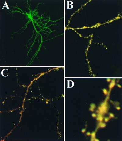
Figure 4.
Expression of YSCS in neurons. Low-magnification images of YSCS fluorescence after excitation of YFP in a neuron (A) from an organotypic hippocampal culture. Images collected during CFP excitation reveal the occurrence of both donor (green) and FRET-based fluorescence (red) throughout the dendritic processes of neurons in organotypic (B) and dissociated (C) hippocampal preparations. Enlargement of a dendrite in slice culture (D) demonstrates that YSCS is concentrated in spine heads.

