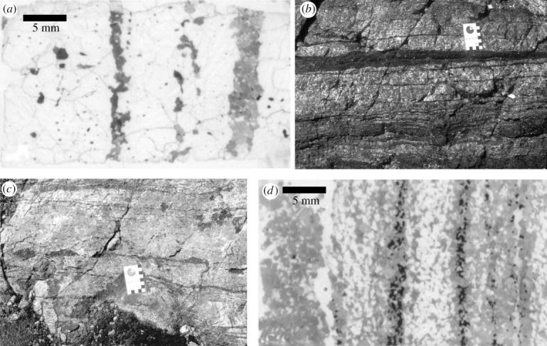
Figure 4.

(a) Scanned full thin section photomicrograph of finely (millimetre-scale) banded quartz and pyroxene rock (sample AK 34). Dark bands are comprised of pyroxene and the white is quartz. (b) Parallel-sided centimetre-thick band of pyroxenite directly below the scale card (in centimetres). (c) Directly above the scale card (in centimetres) shows a trail of pyroxenite boudins parallel to main foiliation. (d) Scanned full thin section photomicrograph showing bands of pyroxene (grey), quartz (white), and magnetite (black). This is sample AK 12, which is shown on figure 2, and also used for U/Pb zircon geochronology.
