
Figure 8.
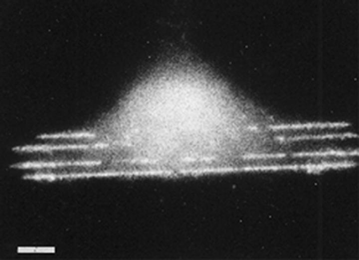
Previously published image (from Wojciak-Stothard et al. 1996) of a macrophage growing on 100 nm deep 5 μm wide grooves fabricated in silica. Fluorescent rhodamine–phalloidin stained. Note lines of actin corresponding to the grooves. Scale bar 5 μm.