
Figure 1.
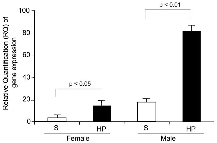
TLR transcripts in the Kupffer cells from male and female mice before and after hypoxia. Male and female mice were subjected to hypoxia (HP) or sham hypoxia (S) for 1.5 h and were killed 1 h after their return to room air. Their livers were removed for Kupffer cell isolation, and Kupffer cell RNA was isolated. The mRNAs of 10 ng were analyzed by real-time PCR using TLR-4–specific primer pairs. The primers of 18S mRNA were included in each reaction as an internal control to normalize relative quantities of TLR. The results are presented in RQ value, which displays the relative level of gene expression in the test samples. This represents 4 identical experiments presented as mean ± SE. P values are shown between 2 indicated groups.