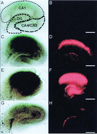
Figure 3.
Influence of OGD and/or thrombin on neuronal survival in organotypic hippocampal slice cultures. Transmission images (Left) and the uptake of fluorescent PI (Right) are shown. Neuronal cell damage can be recognized by dark slice areas and red PI fluorescence in the transmission and fluorescence images, respectively. A control slice is shown in A and B. Transient OGD (30 min) was followed by a pronounced neuronal damage in the CA1 region 24 h later (C and D). Exposure to a relatively high thrombin concentration (500 nM, 1-h incubation) in the presence of oxygen and glucose caused even greater damage (E and F). In contrast, thrombin at a concentration as low as 50 pM, given immediately before and during the OGD, induced significant neuroprotection (G and H). (Bars = 500 μm.)

