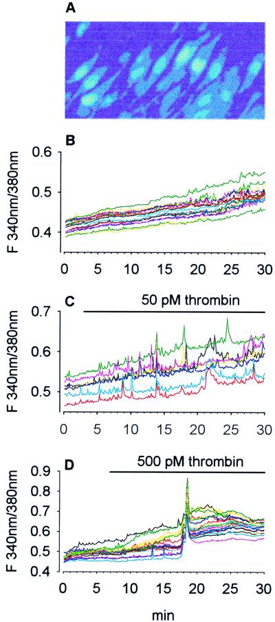
Figure 5.
Thrombin-induced intracellular Ca2+ elevations in hippocampal CA1 neurons. After loading of organotypic slice cultures with fura-2, individual pyramidal cells in the CA1 region can be identified easily (A). Only their soma was used for Ca2+ measurements. Slices were exposed either to a thrombin-free (B) or thrombin-containing (C and D) perfusion medium. Both thrombin concentrations raised [Ca2+]i albeit with different characteristics. Single or repetitive Ca2+ spikes were observed with 50 pM thrombin, whereas a sustained Ca2+ elevation was induced by 500 pM. For each agonist concentration, a typical experiment (of 11) is shown. Each line illustrates the response of an individual cell within the same slice. Fluorescence images were captured every 8 s.

