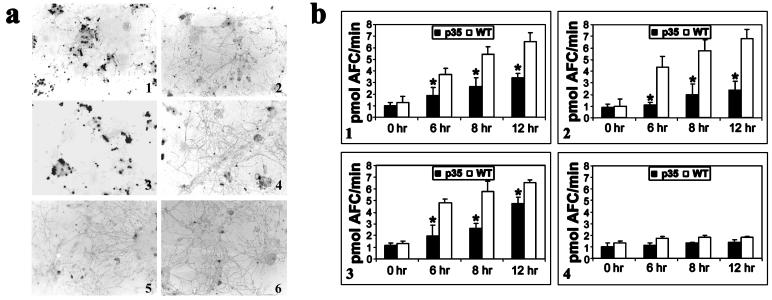
Figure 2.
(a) TUNEL staining of cells 12 h after treatment with staurosporine or growth in lowered extracellular K+. (1 and 3) CGCs isolated from wild-type animals 12 h after growth in low K+-containing medium (5 mM) or 0.5 μM staurosporine, respectively. (2 and 4) CGCs isolated from p35 transgenics after growth in either lowered extracellular K+ or staurosporine, respectively. (5 and 6) CGCs from untreated wild-type and p35 transgenics, respectively. (b) Time course of caspase-3 and -8 activities in cytosolic protein extracts isolated from wild-type (WT) vs. p35 transgenic (p35) CGCs after growth in either low K+ or staurosporine. (1 and 3) Caspase-3 activity assayed fluorometrically by the cleavage of acetylated DEVD-AFC substrate in extracts from CGCs induced to undergo apoptosis by lowered K+ or staurosporine, respectively. (2 and 4) Caspase-8 activity as measured by cleavage of fluorescent IETD-AFC substrate in extracts from CGCs treated with either lowered K+ or staurosporine, respectively. Values represent the means ± SD from three experiments. (*, P < 0.01.)

