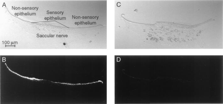
Figure 3.
Immunolabeling of saccular cryosections with the enriched scFv. To characterize the enriched clone, we immunolabeled cryosections of the bullfrog's sacculus with the soluble scFv and examined them by confocal microscopy. (A) A differential-interference–contrast image reveals the components of the saccular epithelium and the underlying saccular nerve. (B) Immunolabeling of the cryosection depicted in A indicates that the antibody fragment recognizes an antigen in the nonsensory epithelium and to a lesser extent in the sensory epithelium of the sacculus. No labeling is seen in the saccular nerve and associated connective tissue. (C) The components of the sacculus are apparent in a second, adjacent cryosection. (D) Omission of the scFv eliminates the intense labeling of the sensory and nonsensory epithelia.

