
Fig. 5. Immunostaining of NIH-3T3 cells expressing human TBX5.
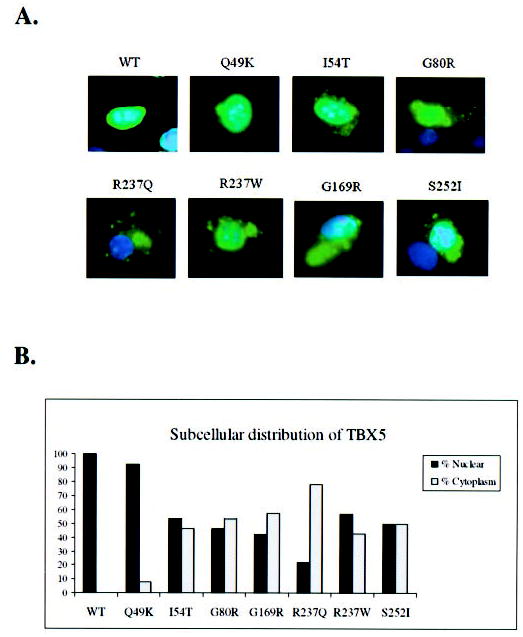
A, WT, cell transfected with the FLAG-tagged wild type TBX5 construct; Q49K, I54T, G80R, R237Q, R237W, G169R, and S252I represent cells over-expressing various mutant TBX5 proteins. Transfected cells were immunostained with anti-FLAG (green) for TBX5 and the nucleus was stained with DAPI (blue). No detectable immunofluorescence staining was observed in non-transfected cells. Note that wild type TBX5 is completely localized into the nucleus, whereas TBX5 proteins with various missense mutations are distributed in both the nucleus and cytoplasm. B, percentage of nuclear versus cytoplasm distribution of TBX5 for wild type (WT) and mutant (Q49K, I54T, G80R, G169R, R237Q, R237W, and S252I). The data were based on the images in panel A.