Figure 3.
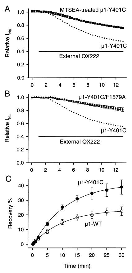
μ1-Y401C creates an access pathway for external QX222 to reach the internal binding site. (A) Effects of MTSEA modification in the outer vestibule on block by external QX222 in μ1-Y401C. MTSEA modification was done by bath application of 2.5 mM MTSEA for 3–5 min and verified by irreversibility of current reduction after washout for at least 5 min before external application of QX222. (B) Effects of mutation of the putative local anesthetic binding site (μ1-F1579) on block by externally applied QX222 in μ1-Y401C. For this, actions of externally applied QX222 were studied for the double mutant, μ1-Y401C/F1579A. In A and B, the protocol was the same as that in Fig. 2, and peak currents were normalized to that in the control. Data represent the means ± SEM from four oocytes for MTSEA-bound μ1-Y401C (■) and five for μ1-Y401C/F1579A (▴). In each panel, the mean values (●) of block for μ1-Y401C shown in Fig. 2C were represented to compare the block development. The bar indicates the period during exposure to 500 μM QX222 in the bath solution. (C) Effects of μ1-Y401C on recovery from internally applied QX222 block. QX222 was internally applied by microinjecting 50 nl of a 3 mM QX222 solution into oocytes. Twenty to thirty minutes after microinjection, a 1-Hz train of 20 pulses with 35-ms duration was applied to −10 mV from a holding potential of −100 mV to produce use-dependent block by QX222. Then, recovery from the QX222 block was monitored at the indicated intervals after the end of a train for μ1-WT (○) and μ1-Y401C (●). Peak currents during recovery were normalized by the difference between the peak current during the first pulse of a train and the 10-s recovery test pulse, and plotted as recovery %. The 10-s recovery was assumed to allow recovery from inactivation of unblocked channels but be short enough to prevent QX222 dissociation from blocked channels. The smooth lines are single exponential fits, and data represent the means ± SEM from six oocytes for μ1-WT and five to seven for μ1-Y401C.

