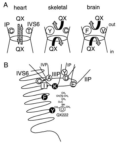
Figure 7.
(A) A model for isoform differences of QX permeation in the Na+ channels. In the heart channel, QX can access to the internal binding site from the outside, and escape to the outside from the cytoplasmic side (16, 21). In skeletal muscle and brain channels, access and escape of QX are limited because of their aromatic residues on the IP-loop (present study), and Cys and Val on IVS6 (16, the present study). (B) Hypothetical pore structure of the Na+ channel composed of four P-loops and IVS6 with respect to access and binding of local anesthetics. Residues shown by open circles (D400, Y401, E755, A1529, and C1572; numbering of μ1) represent the amino acids affecting QX access from the outside (refs. 16 and 27, and the present study) and closed circles (K1237, F1579, and Y1586) affecting local anesthetic binding (15, 17, 27). Ile in square (I1575) appears to affect both access and binding of local anesthetics (15). The DEKA locus (Asp-Glu-Lys-Ala) forms the selectivity ring, shown here as an oval (23).

