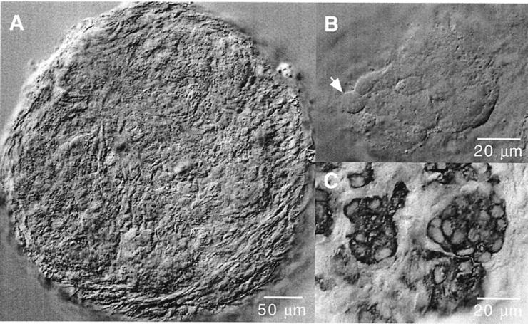
Figure 1.
Morphological appearance of cells in thin slices of rat carotid body. (A) Low-magnification view of a slice a few hours after cutting. (B) Typical cell aggregate (glomerulus) in a carotid-body slice maintained for 48 h in a CO2 incubator. Individual cells, like the one indicated by the arrow, can be clearly differentiated. (C) Carotid-body slice stained with antibodies anti-tyrosine hydroxylase. Note the typical appearance of glomus cells with large nuclei and the organization of the carotid body in glomeruli.

