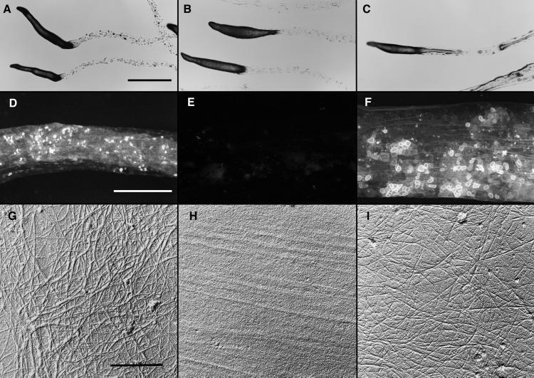
Figure 4.
Slime trails produced by slugs of wild-type, dcsA−, and the dcsA rescued strains. Migrating slugs were phototactically directed to migrate across 2% water agar dishes. They were photographed in place (A–C) or the trails were collected onto coverslips (D–F) or electron microscope specimen grids (G–I). (A–C) All strains formed normal-appearing slugs that migrated phototactically and left behind slime trails. (Bar = 1 mm.) (D–F) The trails left behind wild-type slugs (D), and those of the rescued strain TL128 (F) fluoresced brightly whereas there was only background fluorescence from the trails left behind the dcsA− strain DG1128 (E). (Bar = 100 nm.) (G–I) Slime trails collected on electron microscope specimen grids were treated with Proteinase K (to remove obscuring proteins) and shadowed unidirectionally from 17° with Pt/C and with C from 85°. Microfibrils were seen clearly in the trails left by the wild type (G) and rescued strain, TL128 (I). Microfibrils were absent from the trails of the dcsA− strain, DG1128 (H). (Bar = 50 nm.)

