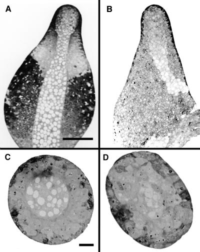
Figure 5.
Comparative sections of developing culminants of wild-type and dcsA− strains. (A and B) Bright-field light micrographs of longitudinal sections of the wild-type (A) and dcsA− strain (B) reveal that the mutant forms the characteristic central core of vacuolized cells but that these do not have the rigid, cellulose-containing walls of normal stalk cells. Moreover, the cellulosic stalk tube is missing such that a functional stalk cannot be made. A first attempt at forming a stalk is apparent at the bottom of the longitudinal section of the mutant (B). (Bar = 50 μm.) (C–D) Bright-field light micrographs of cross-sections of the wild-type (C) and dcsA− strain (D) show the well-defined stalk tube in the wild type (C). A central core of vacuolated cells is present in the mutant, and there is the suggestion of a defining border (D), but the region is not nearly as well defined as in the wild type. (Bar = 10 μm.)

