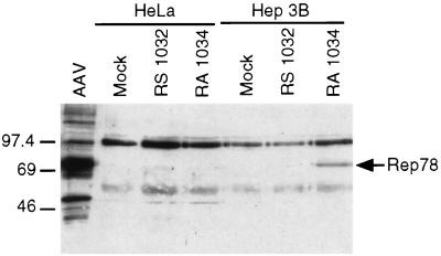
Figure 2.
Western blot analysis of protein extracted from HeLa and Hep3B cells infected with Rep78-expressing Ad vectors. Cells (1.2 × 10⁶) were infected with HDRA1034 or HDRS1032 at an moi of 50 bfu/cell. An AAV-infected 293 cell extract was included in the experiment as positive control. Cell extracts equivalent to 3 × 10⁵ cells were separated on 7.5% SDS/PAGE and transferred to nitrocellulose membrane. Rep polypeptides were detected with a polyclonal rabbit antiserum and chemiluminescence kit (ECL; Amersham Pharmacia). Rep78 polypeptide is indicated on the right margin. A cross-reacting protein of slightly lower molecular weight of Rep78 was detected in some experiments.

