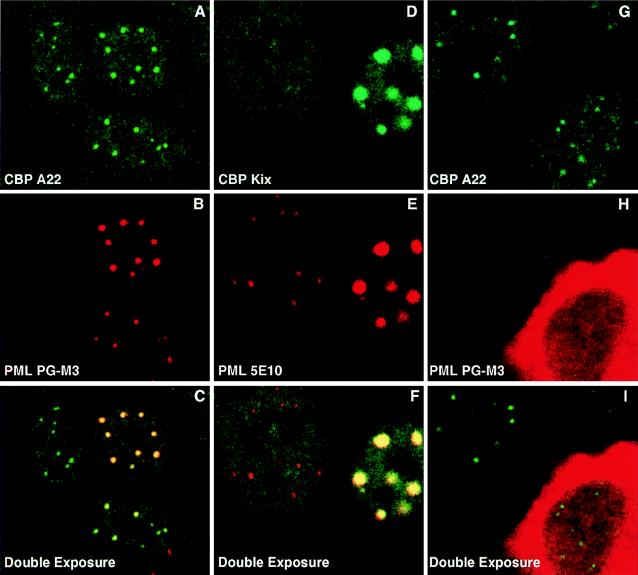
Figure 3.
PML recruits CBP to PODs. (A–I) Double immunofluorescence of CV1 cells (6-well dishes) analyzed in confocal microscopy. Cells were transfected at 70% confluence with 2.5 μg pCMXPML expression vector (A–C); 1 μg of pCMXPML and 2.5 μg pCMXCBPm (mouse) (D–F); and 2.5 μg pSVPMLΔ216-331 (G–I). Primary Abs were used as indicated. Green corresponds to the CBP staining revealed with the FITC-conjugated secondary Ab, red corresponds to the PML staining revealed with the Texas red-conjugated secondary Ab, and the yellow color in the double-exposure image indicates the sites where PML and CBP colocalize.

