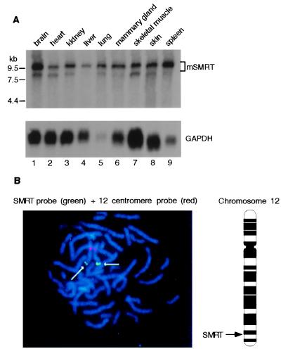
Figure 3.
Tissue distribution and chromosomal localization of SMRT. (A) Poly(A) RNA derived from 20 μg of total RNA from each indicated tissue was subjected to Northern blot analysis. The filter was hybridized with 32P-labeled m-SMRT/PstI fragment (720 bp) (Upper). The relative position of RNA marker (kb) is labeled at the left. The filter was stripped and subsequently reprobed with the glyceraldehyde-3-phosphate dehydrogenase (GAPDH) cDNA for the purpose of loading control (Lower). A 10-kb major transcript was detected in all tissues examined, with relatively high expression in brain, lung, and spleen (lanes 1, 5, and 9). A minor transcript of 8.5 kb in size was present in most tissues. (B) Fluorescence in situ hybridization analysis of h-SMRT is shown with a SMRT-specific probe (green) and a chromosome 12-specific alpha satellite probe that hybridizes to the pericentromeric region (red). The arrows indicate the localization of the SMRT clone at band 12q24. The schematic diagram of chromosome 12 highlights the relative position of SMRT on the chromosome as being close to the 12q24 band. A more defined location is available from genemap 98 (see text).

