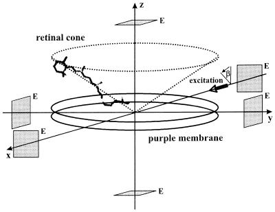
Figure 1.
Geometry of a pm oriented with its membrane normal coincident with the laboratory fixed z axis. The individual retinal chromophores’ transition dipoles form a cone about this axis. The actinic laser pulse propagates along the laboratory fixed x axis with its polarization vector making an angle β with the z axis. The rectangular symbols represent the measuring electrodes (E).

