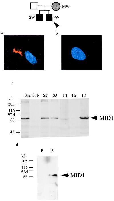
Figure 4.
Four-base pair deletion in a family with Opitz syndrome. (a) Immunofluorescence staining of fibroblasts from a fetus affected with Opitz syndrom (FW) with a MID1-specific antibody. Blue is the DAPI staining of the nuclei. Cytoplasmic clumps were detected with a Cy3-labeled secondary antibody. (b) Immunfluorescence with the same fibroblasts as in A carried out after preincubation of the anti-MID1 antibody with antigenic N-terminal peptides. (c) Cell fractionation of embryonic fibroblasts from a fetus affected with Opitz syndrome. Pellets were resuspended in 2 ml of detergent-containing buffer as in Fig. 1c, and 150 μl of each fraction was separated on a 7.5% polyacrylamide gel, blotted, and probed with affinity-purified anti-MID1 antibody. In the lane of fraction S1a, the same amount of protein (100 μg) was loaded as was used for S1a in Fig. 1c. (d) Western blot analysis of sedimented, reassembled HeLa microtubules in the presence of cytosol of primary embryonic fibroblasts from an OS fetus; 20 μg of the microtubule-containing pellet (lane 1) and the acetone-precipitated, resuspended supernatant (lane 2) were loaded.

