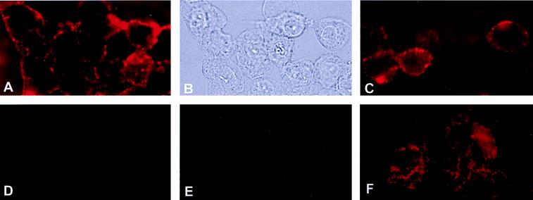
Figure 5.
Immunofluorescence microscopy of ATP-synthase on HUVEC surface. HUVEC were incubated with rabbit polyclonal antiserum raised against the α-subunit of ATP synthase from E. coli as described in Materials and Methods. (A) HUVEC under epi-illumination showing immunofluorescent surface staining for the α-subunit of ATP synthase. (B) Same field of HUVEC under visible light. (C) Human dermal microvascular endothelial cells also showed immunofluorescent surface staining for the α-subunit of ATP synthase. Control experiments were performed with (D) preimmune serum and (E) secondary antibody alone. (F) HUVEC were permeabilized by acetone fixation before adding antibodies for the α-subunit of ATP synthase.

