
Figure 5.
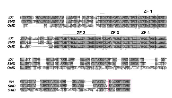
Alignment of entire peptide sequences of ID1, OsID and SbID. Amino acid residues shared by all three peptides are shaded. Each zinc finger is indicated with a light colored box; the putative NLS is shown by a dark bar and the TRDFLG motif is boxed. The boxed area with dotted line shows the C-terminal peptide region used to generate anti-ID1 specific antibody and corresponding sequences in rice and sorghum.