
Fig. 4.
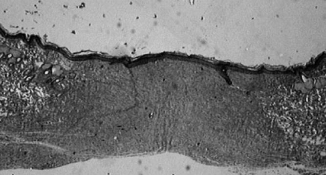
Verhoeff’s staining of wound area and its wound margin in rat treated for 45 days with 1% di-rhamnolipid in eucerin ointment (2× magnification). Evidence of developed fibrosis in wound area with maturing collagen fibers around wound margins. Photograph of histopathology slide created with Issa Version 3.1 software (Vamstec, Vamstec Analysis and Measurement System Technologies, Zagreb, Croatia).