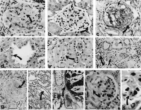
Figure 2.
Kidney histology of adult wild-type, heterozygote (Wt1tmT396/+), and chimeric (Wt1tmT396/+ ↔ +/+) mice. (a) Heterozygote. Glomerulus showing global sclerosis of the glomerular tuft and obliteration of the glomerular capillary bed and urinary filtration space (small arrow) by a dramatic increase in extracellular mesangial matrix (large arrow). The mesangial cells showed increased nuclear size and a prominent nucleolus. Mesangial hypercellularity and podocyte hypertrophy also were evident. The MS was diffuse and affected >90% of glomeruli. (b) Wild type. Glomerulus showing patent glomerular capillaries (small arrow) and urinary filtration space (large arrow) and a normal amount of mesangium and mesangial matrix. (c) Heterozygote. Glomerulus showing focal crescent formation (resulting from proliferation of the Bowmans capsule and macrophages, large arrow) and podocyte hyperplasia (small arrow). (d) Heterozygote. Proximal convoluted tubule showing dilation and microcyst formation. The cysts were lined by hypertrophic tubular epithelial cells with an increased nuclear/cytoplasmic ratio and prominent apically displaced nuclei (arrow). Occasional mitotic figures were evident. (e) Wild type. Proximal convoluted tubule showing normal tubular epithelium with the small luminal space present (arrow) and basal polarization of the nuclear position. (f) Heterozygote showing presence of protein casts in some dilated tubules (arrowhead) and interlobular arteries with medial hyperplasia (small arrow) and interstitial fibrosis (large arrow) secondary to hypertension as a consequence of the diffuse global MS. Pale eosinophilic material and shed epithelial cells also were evident in some cyst lumina. (g) Heterozygote. Interlobular artery showing severe hypertensive damage, including fibrinoid necrosis (arrow), medial hypertrophy and hyperplasia, and loss of the arterial lumen. There was also considerable endothelial swelling and inflammatory infiltrate surrounding many damaged blood vessels. (h) Wild type. Interlobular artery showing small media, thin intima, and patent lumen (arrow). (i) Heterozygote. Electron micrograph showing obliteration of the glomerular tuft (with no capillary bed or glomerular basement membrane) and its replacement by an electron-dense extracellular matrix accumulation (large arrow) containing mesangial cell cytoplasmic processes (small arrows). Podocytes showed loss of foot processes and microvillus transformation of the apical surface. (j) Chimera. Glomerulus showing segmental sclerosis with a normal segment (large arrow) containing patent capillaries and a normal density of mesangial cells, and affected segment (small arrow) with accumulation of mesangial cells, increase in mesangial cell matrix, and obliteration of the glomerular capillary bed. In the majority of glomeruli, the podocytes and the glomerular basement membranes were normal and only occasional podocyte hypertrophy was evident. There was no crescent formation and the blood vessels were normal with no evidence of hypertensive damage. (k) Chimera. Glomerulus showing an apoptotic body (arrow) within the mesangium. [Bars = 60 μm (a–e), 120 μm (f–h), 0.5 μm (i), 60 μm (j), 15 μm (k).]

