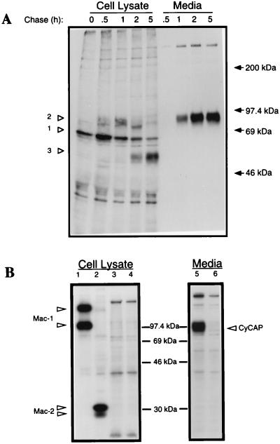
Figure 2.
Biosynthesis and secretion of CyCAP. (A) Pulse–chase analysis of CyCAP in MM55 kidney cells. Cells were pulsed for 10 min with [35S]cysteine and [35S]methionine and chased for the indicated times. CyCAP was immunoprecipitated from cell lysates and media with polyclonal rat serum against CyCAP. Control precipitations with normal rat serum were negative (data not shown). Proteins were resolved by using SDS/7.5% PAGE. Closed arrows represent positions of molecular-mass standards (Rainbow, Amersham Pharmacia). (B) CyCAP secretion from thioglycolate-elicited peritoneal macrophages. PECs were metabolically labeled overnight, and cypC-GST-glutathione agarose was used as an affinity reagent for CyCAP in the absence of CsA (lanes 3 and 5) or in the presence of CsA (lanes 4 and 6). Antibodies to macrophage markers Mac-1 and Mac-2 were used as positive controls (lanes 1 and 2).

