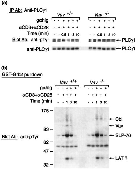
Figure 2.
TCR-induced tyrosine phosphorylation. Immunoblot of cytoplasmic extracts of CD4+ splenic T cells purified from Vav+/+ or Vav−/− mice. Where indicated cells were precoated with anti-CD3 and anti-CD28 antibodies (αCD3 + αCD28) that in some samples were then crosslinked with goat anti-hamster Ig polyclonal antiserum (gαhIg; 100 μg/ml) and samples taken after the indicated time. (a) Samples were immunoprecipitated (IP) with antibodies to PLCγ1, and analyzed by immunoblotting with an antibody to phosphotyrosine (pTyr) and then stripped and reprobed with an antibody to PLCγ1. (b) Proteins binding to a GST–Grb2 fusion protein were isolated from stimulated cell extracts and analyzed by immunoblotting with an anti-pTyr antibody. The identity of individual pTyr-containing proteins was suggested by reprobing the blot with antibodies to Cbl, Vav, and SLP-76 (data not shown). The 36-kDa phosphoprotein is likely to be LAT (38). Sizes are shown in kDa.

