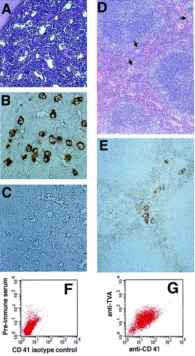
Figure 2.
ALV receptor protein is expressed in murine hematopoietic tissue. Hematoxylin/eosin stains of bone marrow (A) and spleen (D) from a TVA-expressing transgenic mouse show grossly normal tissue. Immunohistochemical staining using an anti-TVA antibody demonstrates TVA expression on megakaryocytes from transgenic mice (B) but not from nontransgenic littermates (C). TVA expression is also seen in splenic megakaryocytes from transgenic mice (E). Independent FACS analysis using isotype controls for anti-CD41 and anti-TVA plotted against forward scatter was used to identify a generous gate that encompasses all cells stained by either antibody. Two-dimensional FACS analysis using isotype controls for anti-CD41 and anti-TVA demonstrates cells congregated in the left lower quadrant of the FACS dot plot (F), with a shift of the population up and to the right after incubation with anti-TVA and anti-CD41 antibodies (G).

